The organ system of human beings (and other animals) which is responsible for the transport of materials inside the body is called circulatory system. The various organs of the circulatory system in humans are: Heart, Arteries, Veins and Capillaries. Blood is also considered a part of the circulatory system. So, the human circulatory system consists of the heart, arteries, veins, capillaries, and blood. In the circulatory system, the heart acts as a pump to push out blood. The arteries, veins and capillaries act as pipes (or tubes) through which the blood flows. These tubes which carry blood are called blood vessels. Thus, there are three types of blood vessels in the human body: arteries, veins and capillaries. We will now describe all the parts of the circulatory system in detail.
Diagram to show the inside structure of human heart.
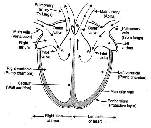
The heart is roughly triangular in shape. It is made of special muscle called cardiac muscle. The size of our heart is about the same as our ‘clenched fisť. The heart has four compartments called ‘chambers’ inside it. The upper two chambers of heart are called atria (singular atrium), and the lower two chambers of heart are called ventricles. The two atria receive blood from the two main veins. And the two ventricles transport blood to the entire body and the lungs. The left atrium is connected to the left ventricle through a valve V1. Similarly, the right atrium is connected to the right ventricle through another valve V2. These valves prevent the backflow of blood into atria when the ventricles contract to pump blood out of the heart to the rest of the body. This is because when the ventricles contract, the valves V1 and V2 close automatically so that the blood may not go back into atria. The job of heart is to pump blood around our body. All the atria and ventricles of the heart contract and relax (expand) at appropriate times and make the heart behave like a pump. Since ventricles have to pump blood into various organs with high pressure, they have thicker walls than atria. A sheath of tissue called ‘pericardium’ protects the muscular heart. The chambers of the heart are separated by a partition called septum.
The arteries, veins and capillaries are a kind of thin pipes (or tubes) through which blood the body. Arteries, veins and capillaries are called blood vessels. Arteries are the thick walled blood vessels which carry blood from the heart to all the parts of the body. Arteries have thick walls blood emerges from the heart under high pressure. Arteries are found in the whole of our body. The main artery (called aorta) is connected to the left ventricle of the heart through a valve V3. The main artery carries oxygenated blood from the left ventricle to all the parts of the body (except the lungs). Another artery called pulmonary artery is connected to the right ventricle of the heart through another valve V4. The pulmonary artery carries deoxygenated blood from the right ventricle to the lungs.
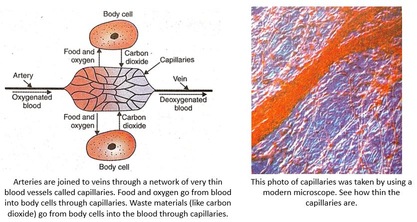
The capillaries are thin walled and extremely narrow tubes or blood vessels which connect arteries to veins. Thus, the capillaries are in-between the arteries and veins. The blood from arteries enters the capillaries in the body. Every living cell of our body is close to a capillary. The walls of capillaries are only one-cell thick. The various dissolved substances (like oxygen, food, etc.) present in blood pass into the body cells through the thin walls of the capillaries. At the same time, the waste substances (like carbon dioxide) formed in the cells enter into capillaries. Thus, the exchange of various materials like oxygen, food, carbon dioxide, etc., between the blood and the body cells takes place through capillaries. The other end of capillaries is joined to some wider tubes called veins. The deoxygenated blood (or dirty blood) coming from the capillaries enters into veins.
Veins are the thin walled blood vessels which carry blood from all the parts of the body back to the heart. Veins do not need thick walls because the blood flowing through them is no longer under high pressure. Veins have valves in them which allow the blood in them to flow in only one direction (towards the heart). The valves prevent the backflow of blood in veins. Veins are also found in the whole of our body. The pulmonary vein is connected to the left atrium of the heart. The pulmonary vein carries oxygenated blood from lungs back to the heart. There is also a main vein (called vena cava). The main vein is connected to the right atrium of the heart. The main vein carries deoxygenated blood from all the parts of the body (except lungs), back to the heart. Please note that the main difference between an artery and a vein is that an artery carries blood from the heart to the body organs whereas a vein carries blood from the body organs back to the heart.
We have just studied that the heart is a kind of pump which pumps blood around our body continuously, without stopping. Actually, heart is not a single pump. Heart is really a double pump. The left side of heart (left atrium and left ventricle) acts as one pump which pumps blood into the whole body, except the lungs. The right side of heart (right atrium and right ventricle) acts as another pump which pumps blood only into the lungs. the left side of heart is completely separated from the right side by a partition called septum. So, the two pumps in the heart work independently. The separation of left and right sides of the heart is necessary to prevent the mixing of the oxygenated blood on the left side with the deoxygenated blood on the right side.
Before we describe the circulation of blood in the human body with the help of a diagram, we should keep the following two points in mind : First that the blood circulates in our body in two forms : oxygenated blood and deoxygenated blood. The blood carrying oxygen in it is called oxygenated blood. We get oxygenated blood in the lungs where the fresh oxygen of air passes into the blood. The blood having no oxygen in it is called deoxygenated blood. The deoxygenated blood is formed in all the organs of the body (except the lungs). This is because when the oxygenated blood passes through the organs of the body, the body cells use up its oxygen and make it deoxygenated. The deoxygenated blood, however, carries carbon dioxide in it (which is produced during respiration in body cells). The second point to remember is that when blood circulates in the body, then it supplies oxygen, digested food and other chemicals (like hormones) to all the cells of the body. It also carries back waste products like carbon dioxide, etc., from the body cells.
The heart beats non-stop all the time. The heart beat is due to the rhythmic contraction and relaxation of the heart muscles which make up the atria and the ventricles. Please note that the two atria (left atrium and right atrium) contract together and relax together. Similarly, the two ventricles (left ventricle and right ventricle) contract together and relax together. The contraction of two atria is immediately followed by the contraction of the two ventricles.
The heart beats (or beating of heart) circulates the blood in the human body. We will now describe the circulation of blood in the human body with the help of a highly simplified diagram
Diagram to show blood circulation in human body.
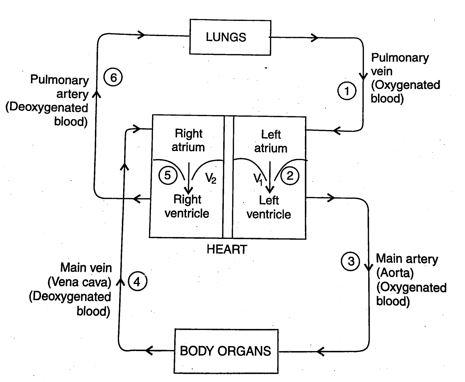
1. When the muscles of all the four chambers of the heart are relaxed, the pulmonary vein brings the oxygenated blood (oxygen-carrying blood) from the lungs into the left atrium of the heart.
2. When the left atrium contracts, the oxygenated blood is pushed into the left ventricle through the valve V1.
3. When the left ventricle contracts, the oxygenated blood is forced into the main artery called ‘aorta’. This main artery then branches into smaller arteries which go into different body organs (except the lungs). The smaller arteries (called arterioles) further branch into capillaries (The smaller arteries and capillaries have not been to keep the diagram simple).
4. The main artery carries blood to all the organs (or parts of the body like head, chest, arms, stomach, intestines, liver, kidney, trunk and legs (except the lungs). When the oxygenated blood passes through the capillaries of the body organs, then it gives oxygen to the body cells. Since the blood loses oxygen here, we say that the blood has been deoxygenated. The blood also gives the digested food and other dissolved materials to the body cells. At the same time, carbon dioxide produced as a waste material during respiration enters into the blood. The deoxygenated blood (carrying carbon dioxide) from the body organs enters into the main vein called vena cava. The main vein carries the deoxygenated blood to the right atrium of the heart.
5. When the right atrium contracts, deoxygenated blood is pushed into the right ventricle valve V2.
6. And when the right ventricle contracts, the deoxygenated blood is pumped into the lungs through the pulmonary artery. In the lungs, deoxygenated blood releases its carbon dioxide and absorbs fresh oxygen from air. So, the blood becomes oxygenated again. This oxygenated blood is again sent to the left atrium of heart by pulmonary vein for circulation in the body.
This whole process is repeated continuously. In this way, the blood keeps on circulating in our body without stopping due to which all the body parts keep on getting oxygen, digested food and other materials all the time. The blood circulation also keeps on removing waste products formed in the cells of the body.
The blood circulatory system in human beings is an example of double circulation. This can be explained as follows : A circulatory system in which the blood travels twice through the heart in one complete cycle of the body is called double circulation. In the human circulatory system the pathway of blood from the heart to the lungs and back to the heart is called pulmonary circulation; and the pathway of blood from the heart to the rest of the body and back to the heart is called the systemic circulation. These two types of circulation taken together make double circulation.
The animals such as mammals (including human beings), and birds have four-chambered heart (which consists of two atria and two ventricles). In a four-chambered heart, the left side and right side of the heart are completely separated to prevent the oxygenated blood from mixing with deoxygenated blood. Such a separation allows a highly efficient supply of oxygen to the body cells which is necessary for producing a lot of energy. This energy is useful in warm-blooded animals (like mammals and birds) which have high energy needs because they constantly require energy to maintain their body temperature. All the animals having four-chambered hearts have double circulation in which the blood passes through the heart ‘twice’ in one complete cycle of the body.
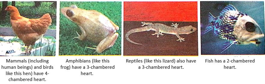
The animals such as amphibians and many reptiles are cold-blooded animals whose body temperature depends on the temperature in the environment. They do not need energy to maintain their body temperature and hence their requirement of energy is less. The amphibians (like frogs) and reptiles (like lizards) have a three-chambered heart (which consists of two atria and one ventricle). Due to incomplete division within their heart, the oxygenated and deoxygenated bloods mix to some extent in amphibians and reptiles. This reduces the production of energy. The amphibians and reptiles have, however, a double circulation that delivers blood to the lungs and the rest of the body, respectively.
The fish has a two-chambered heart (which consists of one atrium and one ventricle). The fish does not have lungs, it has gills to oxygenate blood. In a fish, the heart pumps deoxygenated blood to the gills. Oxygenation of blood takes place in the gills. The oxygenated blood from the gills is supplied to the body parts of the fish where oxygen is utilised and carbon dioxide enters into it making it deoxygenated. This deoxygenated blood returns to the heart to be pumped into gills again. The flow of blood in a fish is called single circulation because the blood passes through the heart of fish only once in one complete cycle of the body.
Heart Beats
Contents
- 1 Heart Beats
- 2 Pulse
- 3 Blood Pressure
- 4 How to Measure Blood Pressure
- 5 How do Food and Oxygen Reach Body Cells
- 6 🫀 Main Components of the Circulatory System
- 7 🔄 Types of Circulation
- 8 ⚙️ Working of the Heart (Simplified Flow Path)
- 9 🔬 Functions of the Circulatory System
- 10 ❗ Common Circulatory System Diseases:
- 11 🧠 Interesting Facts:
The heart pumps blood into our arteries by contracting. When the heart contracts, it becomes smaller in size and pushes the blood into main artery with a great force. Then the heart relaxes (comes back to its original size) and gets filled up with blood from pulmonary vein. In this way, the heart keeps on contracting and relaxing again and again to pump blood into the body continuously. One complete contraction and relaxation of the heart is called a heart beat. The heart usually beats about 70 to 72 times in a minute when we are resting. This means that the heart pumps out blood to the arteries about 70 to 72 times per minute.
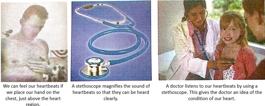
We can feel our heart beats if we place our hand on the chest just above the heart region. A doctor listens to our heart beats by using an apparatus called stethoscope. The stethoscope magnifies the sound of heart beats so that the doctor can hear the heart beats clearly.
Though the average number of heart beats of a person at rest is about 70 to 72 per minute but the number of heart beats increases too much after a physical exercise or when a person is excited. For example, if we count our heart beats after running for a while, we will find it to be more than 100 per minute. The heart beats faster during and after an exercise because the body needs more energy under these conditions. The faster beating of heart pumps blood more rapidly to the body organs which supplies more oxygen to the body cells for rapid respiration to produce more energy. The heart beats can be counted easily by counting the pulse.
Pulse
Every time the heart beats, blood is forced into arteries. This blood makes the arteries expand a little. The expansion of an artery each time the blood is forced into it, is called pulse. Each heartbeat generates one pulse in the arteries, so the pulse rate of a person is equal to the number of heartbeats per minute. Since the heart beats about 70 to 72 times per minute, therefore, the pulse rate of an adult person while resting is 70 to 72 per minute. Thus, the pulse rate is the same as the heart rate. Just like heartbeats, the pulse rate of a person is higher after a physical exercise or when a person is excited.
Most of our arteries lie deep inside our body and hence cannot be used to feel the pulse. But at some places in our body like the wrist, temple and neck, the arteries are close to the surface of skin and pass over bones. So, we can feel the pulse at wrist, temple and neck by pressing the artery lightly with our finger tips. The pulse is traditionally taken above the wrist. We can feel our own find the pulse rate as follows:
Feeling the pulse in the wrist.
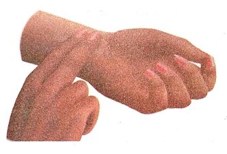
The pulse can be felt with fingers placed gently on arteries at the wrist. We place the first two fingers (index finger
ace the first two fingers (index finger and middle finger of our right hand on the inner side of our left wrist and press it gently. We will feel some waves touching our fingers. These waves are the pulse. We can count the number of such waves (or thumpings) in one minute by using a watch. This will give us the pulse rate (per minute).
We usually see the doctor taking the pulse rate of a patient by keeping his fingers on the wrist of the patient and at the same time looking into his watch. Doctors can tell by counting the pulse rate and listening to heartbeats whether a person is well or not. This is because the pulse rate and heartbeats change according to the condition of our heart.
Blood Pressure
The pressure at which blood is pumped around the body by the heart is called blood pressure. The blood pressure of a person is always expressed in the form of two values called ‘systolic pressure’ and ‘diastolic pressure‘. In order to understand this, we should first know the meaning of ‘systole’ and ‘diastole’. The phase of the heart beat when the heart contracts and pumps the blood into arteries is called ‘systole’. And the phase of heart beat when the heart relaxes (or expands) and allows the chambers to fill with blood is called ‘diastole’.
The maximum pressure at which the blood leaves the heart through the main artery (aorta) during contraction phase, is called the systolic pressure. This high pressure in the main artery maintains a steady flow of blood in all the arteries towards the capillaries. The minimum pressure in the arteries during the relaxation phase of heart is called the diastolic pressure. The value of diastolic pressure is always lower than that of the systolic pressure. The blood pressure of a person is expressed in terms of millimetres of mercury (which is written as mm Hg). The normal blood pressure values are :
Systolic pressure : 120 mm Hg
Diastolic pressure : 80 mm Hg
This is usually written as 120/80
The blood pressure values vary from person to person and from time to time. They also vary with age. For example, a young person may have blood pressure of 110/75 but at the age of 60 years it could be 145/90. High blood pressure is called hypertension. High blood pressure is caused by the constriction (narrowing) of very small arteries (called arterioles) which results in increased resistance to blood flow. Very high blood pressure can lead to rupture of an artery and internal bleeding.
How to Measure Blood Pressure
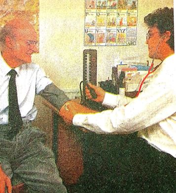
Blood pressure is measured by using an instrument called sphygmomanometer. Two readings of blood pressure are taken : systolic pressure (when the heart is contracting and pumping out blood), ana diastolic pressure (when the heart relaxes and fills with blood). The various steps in measuring the blood pressure of a person are as follows:
(i) A rubber cuff (which is a flat rubber tube) is wrapped around the person’s arm. The rubber cuff is inflated by pumping air into it to give a pressure of about 200 mm Hg to the brachial artery (which runs down the arm). This pressure can be seen on the scale of the instrument sphygmomanometer. If a stethoscope is now placed on the artery of the arm, no sound is heard through it.
Measuring of blood pressure by using a mercury sphygmomanometer.
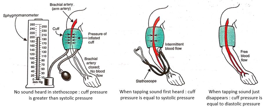
(ii) With stethoscope still placed on artery, the cuff pressure is reduced gradually by deflating it. The cuff pressure when the heart beat is first heard as a soft tapping sound through the stethoscope gives us the systolic pressure.
(iii) The cuff pressure is reduced further by deflating it more and more. The cuff pressure when the tapping sound in stethoscope just disappears, gives us the diastolic pressure.
The above observations can be explained as follows: When a high pressure of about 200 mm Hg is applied to the arm by the cuff, then the brachial artery gets closed fully and hence no blood flows in it. Since no blood flows in the brachial artery at this stage, therefore, no tapping sound is heard in the stethoscope. When the cuff pressure is reduced and becomes equal to the systolic pressure, then the brachial artery opens up slightly and there is an intermittent blood flow in it due to which a soft tapping sound just begins to be heard in the stethoscope. And finally, when the cuff pressure is reduced further and it becomes equal to diastolic pressure, then the brachial artery opens up fully, the blood flow in it is fully restored and hence the tapping sound just disappears.
A doctor measuring the blood pressure of a patient by using a sphygmomanometer.
How do Food and Oxygen Reach Body Cells
We have studied that blood carries food and oxygen around the body. But blood never comes in contact with body cells. So, how do food and oxygen get from the blood to the body cells where they are needed ? This happens with the help of plasma which leaks from the blood capillaries around the body cells. This plasma which leaks out from the blood capillaries is called tissue fluid. We can now say that : The liquid from the blood which is forced out through the capillary walls and moves between all the body cells (providing them with food and oxygen, and removing carbon dioxide) is called tissue fluid.
Actually, the walls of blood capillaries are very thin. So, when blood flows through the capillaries, a liquid called tissue fluid leaks from the blood capillaries and goes into tiny spaces between the various body cells in the tissues. The tissue fluid carries food and oxygen from the blood to the cells, and picks up their waste products like carbon dioxide. After doing its job, most of the tissue fluid seeps back into blood capillaries. The remaining tissue fluid carrying large protein molecules, digested fat, germs from the cells and fragments of dead cells, enters into another type of tiny tubes called lymph capillaries and it becomes lymph. This lymph (alongwith its contents) is returned to the blood by another type of transport system in the human body called lymphatic system.
You can write your questions and suggestions to us in the comment box given below.
Thank you
The human circulatory system is a vital organ system responsible for the transport of blood, oxygen, nutrients, hormones, and waste products throughout the body. It plays a key role in maintaining homeostasis.
🫀 Main Components of the Circulatory System
1. Heart ❤️
-
A muscular organ located slightly to the left of the chest.
-
Pumps blood throughout the body via rhythmic contractions.
-
Divided into four chambers:
-
Right Atrium
-
Right Ventricle
-
Left Atrium
-
Left Ventricle
-
2. Blood Vessels 🩸
There are three major types:
-
Arteries: Carry oxygenated blood away from the heart (except pulmonary artery).
-
Veins: Carry deoxygenated blood toward the heart (except pulmonary vein).
-
Capillaries: Tiny blood vessels where exchange of gases, nutrients, and waste occurs.
3. Blood 🧬
-
Composed of plasma (liquid part), red blood cells (RBCs), white blood cells (WBCs), and platelets.
-
Functions:
-
RBCs carry oxygen (via hemoglobin).
-
WBCs fight infection.
-
Platelets help in blood clotting.
-
Plasma transports nutrients and hormones.
-
🔄 Types of Circulation
1. Pulmonary Circulation
-
Carries deoxygenated blood from the heart to the lungs and brings back oxygenated blood to the heart.
➡️ Right Ventricle → Lungs → Left Atrium
2. Systemic Circulation
-
Carries oxygenated blood from the heart to rest of the body and returns deoxygenated blood back.
➡️ Left Ventricle → Body → Right Atrium
3. Coronary Circulation
-
Supplies blood to the heart muscle itself.
4. Portal Circulation
-
A part of systemic circulation where blood from digestive organs passes through the liver before entering general circulation.
⚙️ Working of the Heart (Simplified Flow Path)
-
Body → Right Atrium (via superior/inferior vena cava)
-
Right Atrium → Right Ventricle
-
Right Ventricle → Lungs (via pulmonary artery)
-
Lungs → Left Atrium (via pulmonary vein)
-
Left Atrium → Left Ventricle
-
Left Ventricle → Body (via aorta)
🌀 This cycle repeats with every heartbeat (~72 times/min in a healthy adult).
🔬 Functions of the Circulatory System
-
💨 Transport: Oxygen, carbon dioxide, nutrients, hormones.
-
🛡️ Protection: WBCs defend against infections, clotting prevents blood loss.
-
🌡️ Regulation: Maintains body temperature, pH, and fluid balance.
❗ Common Circulatory System Diseases:
| Disease | Description |
|---|---|
| Hypertension | High blood pressure |
| Atherosclerosis | Fat deposits in arteries |
| Heart Attack (Myocardial Infarction) | Blockage in coronary arteries |
| Stroke | Interrupted blood flow to brain |
| Anemia | Low RBCs or hemoglobin |
🧠 Interesting Facts:
-
The heart beats over 100,000 times a day.
-
Blood travels about 12,000 miles in one day.
-
The body has about 5-6 liters of blood.
Would you like a diagram of the circulatory system or this explanation in Hindi?